Peripheral Angiogram

Diagnostic peripheral angiography is a minimally invasive imaging test that uses X-rays and a special dye to see inside the arteries that carry blood from the heart to the extremities—the arms, legs, hands, and feet.
Interventional cardiovascular specialist Dr. Shawn Howell uses peripheral angiograms, also called arteriograms, to locate blockages in arteries if she suspects you have peripheral artery disease (PAD).
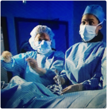
The test is performed on an outpatient basis in a cardiac catheterization laboratory. An IV containing a sedative to help you relax will be given, as well as a local anesthetic to numb the location of the needle puncture. Dr. Howell will then insert a catheter into the artery, injecting a small amount of dye so that imaging of your narrowed or blocked arteries is clear. X-ray images will then be taken.
You may feel pressure during your procedure, but it is not painful. The dye may cause a hot or flushed sensation. The procedure typically takes about an hour, but may take longer. Recovery after a peripheral angiogram usually takes six to eight hours.
After Dr. Howell reviews your test results, she will go over your treatments for narrowed or blocked arteries, which may include peripheral angioplasty. She will take time to ensure you understand your treatment options and help you choose what is best for you.
Preparing for Peripheral Angiography
Before the procedure, it is important to report allergies to X-ray dye and shellfish (both have iodine). Patients who are allergic to either will be given medications prior to the procedure to prevent a reaction. No food should be eaten for 8 to 12 hours before the test.
When you have narrowed or blocked arteries, you need a board-certified vascular specialist with a proven track record of optimal patient outcomes. Call (202) 466-3000 to schedule an appointment with Dr. Howell or use our online appointment request form today.